Scheduled biopsy, unexpected bleeding
The video requires an internet connection.
History
A 74-year-old female patient, with chronic HBV infection, diabetes mellitus and a history of hysterectomy, known with a neglected cystic pancreatic lesion (mucinous cystadenoma), in whom the latest CT evaluation shows a pancreatic tumour with a cystic component, most likely a cystadenocarcinoma, is admitted for EUS-FNB to obtain a definitive diagnosis.
Clinical & biological
Clinical examination shows upper abdominal tenderness and splenomegaly.
Laboratory tests reveal hypochromic microcytic iron-deficiency anaemia, thrombocytopenia, an inflammatory syndrome and CA 19-9 > 1000 U/mL.
Imaging
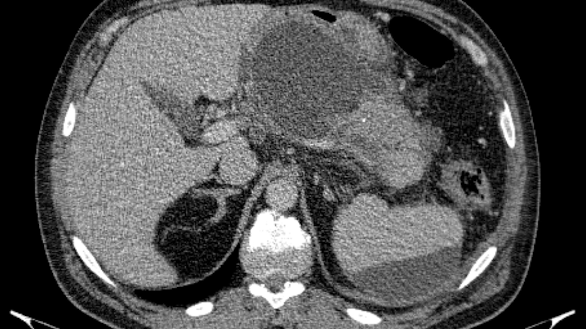
CT: cystic-solid pancreatic tumour invading the SMV, the preconfluent SV and the PV trunk; secondary portal hypertension; non-cirrhotic liver; ascites, peritoneal M1 under observation.
EUS: bleeding from a variceal channel, with interruption of the procedure.
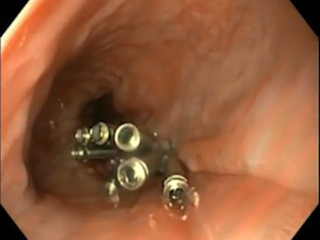
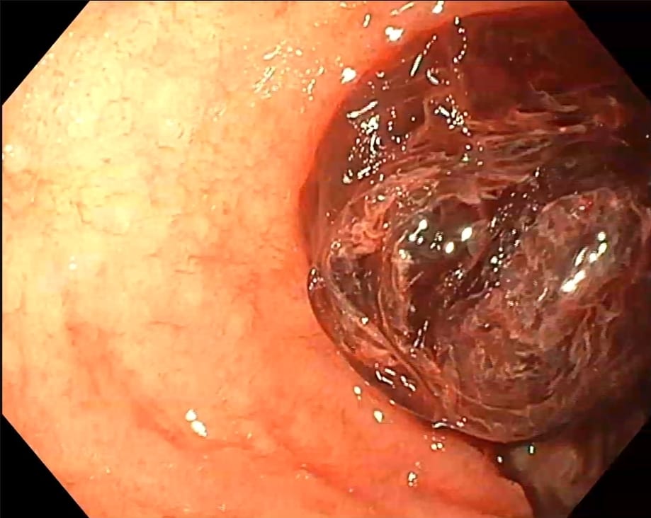
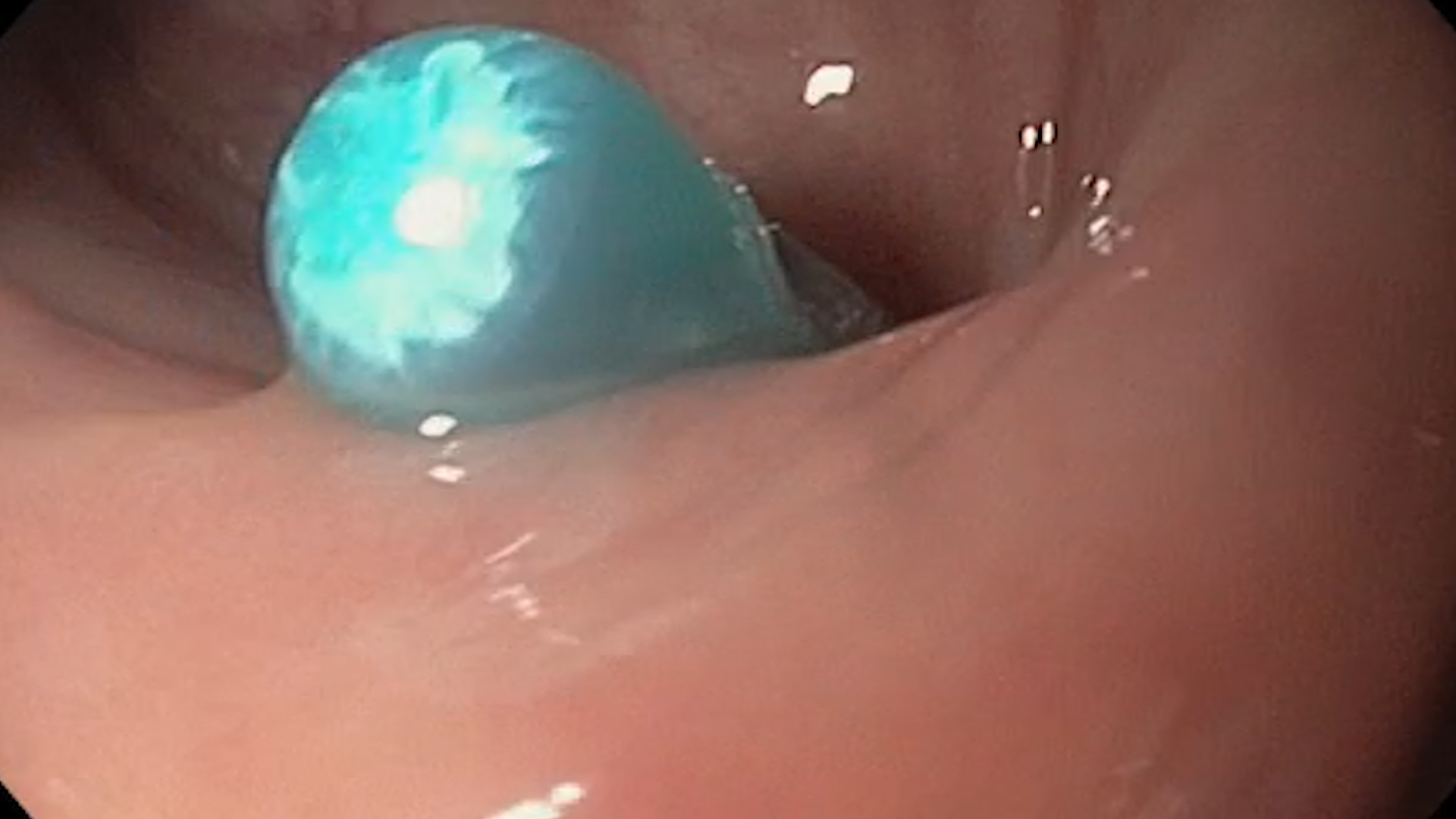
EGD: large oesophageal varices with active bleeding; endoscopic band ligation (7 bands on 3 variceal columns), with bleeding control at the end of the procedure (Figures 1-5, Video 1).
Figure legends:
Photo 1 – Large oesophageal varices seen through the band-ligator cap.
Photo 2 – Active variceal bleeding, with the oesophageal lumen filled with blood.
Photo 3 – Oesophageal variceal columns and blood in the lumen during ligation.
Photo 4 – Variceal column suctioned into the cap before band release.
Photo 5 – Post-ligation appearance: varix strangulated by the band ("pseudopolyp").
Diagnosis
Discussion
• Mucinous cystadenoma has a known malignant potential; in this patient, lack of surveillance most likely allowed progression to cystadenocarcinoma, diagnosed only at an advanced stage, with vascular and peritoneal invasion.
• In this case, portal hypertension is exclusively prehepatic, caused by tumour invasion of the SMV, SV and PV, in a non-cirrhotic liver.
• Splenomegaly and thrombocytopenia may predict the presence of gastro-oesophageal collaterals, so an EGD before endoscopic ultrasound (EUS) may be justified.
• Haemostasis was achieved by immediate endoscopic band ligation, applied to all 3 variceal columns.
• Portal trunk invasion complicates TIPS as a rescue option, making variceal eradication by repeated ligation the only real protection against rebleeding.
Conclusions
• Mucinous cystadenoma requires rigorous periodic surveillance; neglecting it may lead to the diagnosis of an advanced cystadenocarcinoma.
• Tumour-related portal hypertension can turn a diagnostic procedure into a haemorrhagic emergency.
• Prompt endoscopic band ligation is life-saving, and variceal eradication is essential for secondary prophylaxis, as beta-blocker therapy is not effective in tumour-related portal hypertension.
References
- European Study Group on Cystic Tumours of the Pancreas. European evidence-based guidelines on pancreatic cystic neoplasms. Gut. 2018;67(5):789-804. doi:10.1136/gutjnl-2018-316027
- Polkowski M, Jenssen C, Kaye P, et al. Technical aspects of endoscopic ultrasound (EUS)-guided sampling in gastroenterology: European Society of Gastrointestinal Endoscopy (ESGE) Technical Guideline - March 2017. Endoscopy. 2017;49(10):989-1006. doi:10.1055/s-0043-119219
- de Franchis R, Bosch J, Garcia-Tsao G, et al. Baveno VII - Renewing consensus in portal hypertension. J Hepatol. 2022;76(4):959-974. doi:10.1016/j.jhep.2021.12.022
- Gralnek IM, Camus Duboc M, Garcia-Pagan JC, et al. Endoscopic diagnosis and management of esophagogastric variceal hemorrhage: European Society of Gastrointestinal Endoscopy (ESGE) Guideline. Endoscopy. 2022;54(11):1094-1120. doi:10.1055/a-1939-4887