Step-up management of recurrent walled-off pancreatic necrosis
The video requires an internet connection.
History
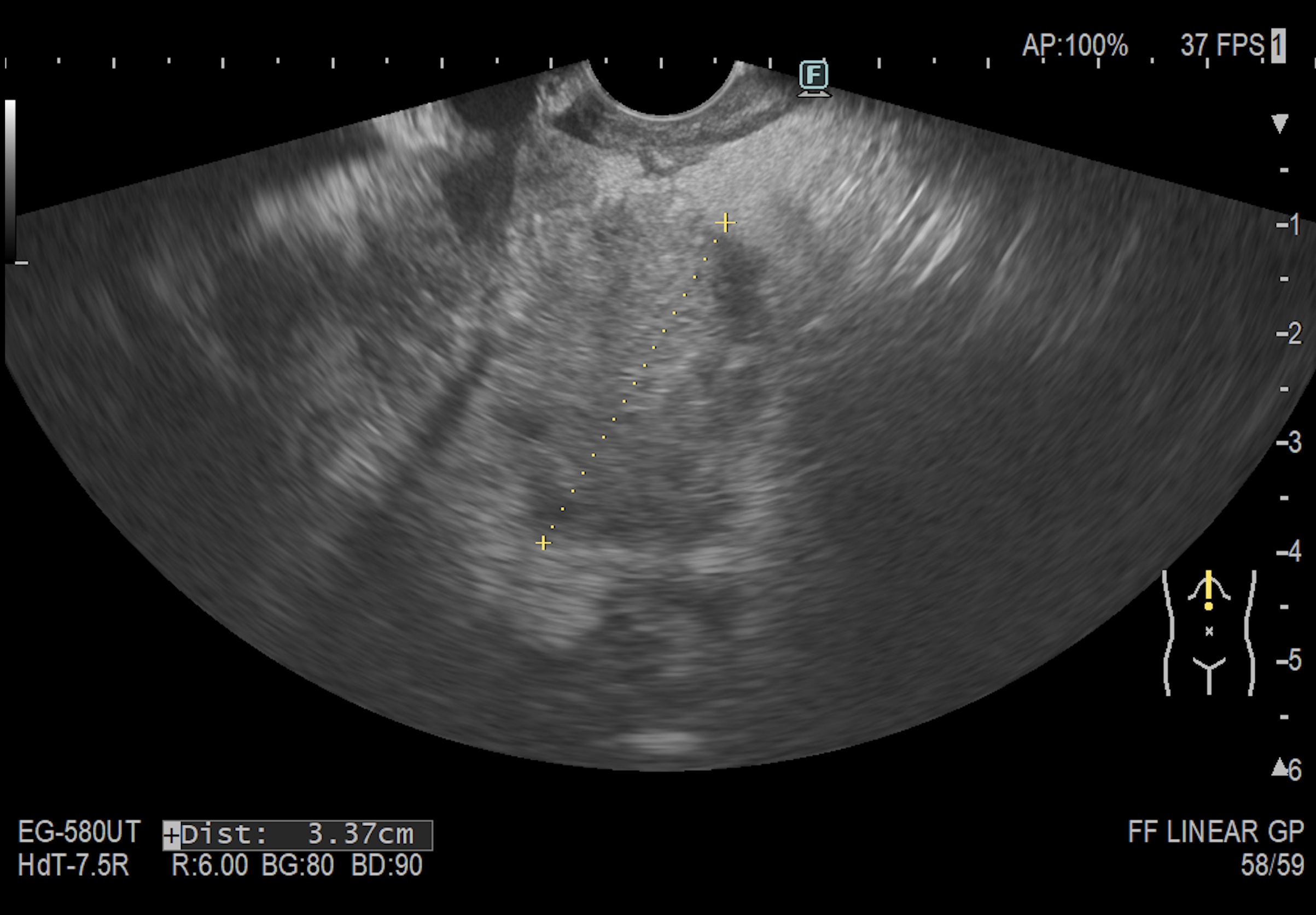
A 38-year-old man with a history of recent alcohol-related acute pancreatitis complicated by a large pancreatic fluid collection, initially measuring approximately 14 cm. The collection was initially partially drained using plastic stents. The clinical course was complicated by prolonged fever, requiring antibiotic therapy with intravenous meropenem and vancomycin. Subsequently, EUS-guided transmural drainage was performed using a lumen-apposing metal stent (Hot AXIOS), followed by five sessions of endoscopic necrosectomy. After four weeks, the Hot AXIOS stent was removed. Following stent removal, the patient initially showed clinical and radiological improvement, with a residual collection measuring 40 × 53 × 45 mm.
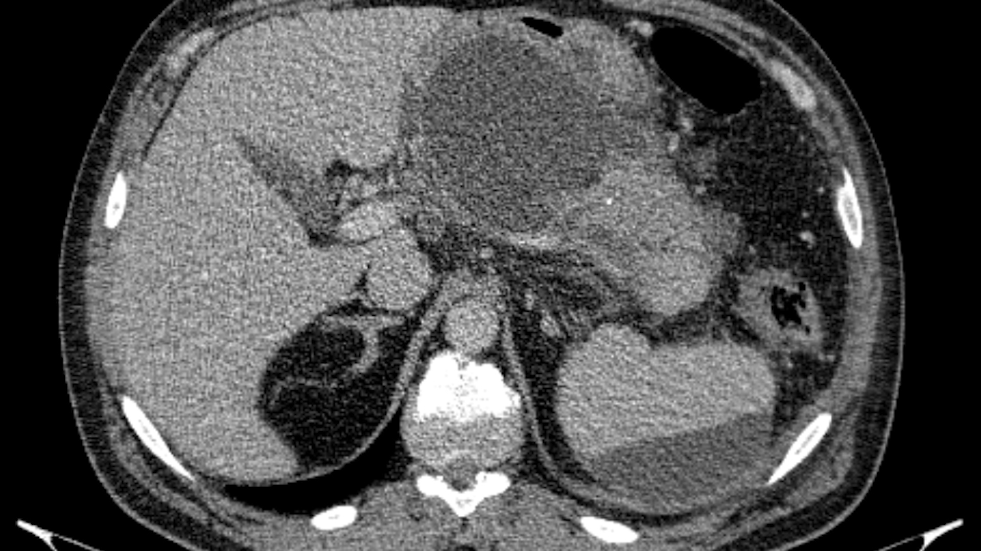
Approximately two weeks later, he was readmitted because of recurrent fever and abdominal pain. CT demonstrated significant enlargement of the collection, inflammatory changes involving the adjacent gastric wall, and suspected focal thrombosis of the splenic vein.
The case was discussed within a multidisciplinary team, and surgical management was subsequently decided.
Relevant history included active smoking, chronic alcohol consumption with recent abstinence, and previous inguinal hernia repair during childhood.
The patient had also experienced approximately 15 kg of weight loss following the episode of acute pancreatitis.
Clinical & biological
On admission, the patient was conscious, cooperative, afebrile, hemodynamically and respiratory stable. The abdomen was soft and non-tender, with preserved bowel movements and urine output.
Laboratory findings showed:
• mild anemia: hemoglobin 12.2 g/dL;
• mild thrombocytopenia;
• elevated C-reactive protein: 10.3 mg/dL;
• low procalcitonin: 0.086 ng/mL.
Imaging
Contrast-enhanced CT
Compared with the CT performed after metallic stent removal, CT demonstrated a significant increase in the size of the pancreatic collection, from 40 × 53 × 45 mm to 62 × 85 × 58 mm. The collection was in close contact with the lesser gastric curvature and caused marked reactive thickening of the gastric wall, predominantly due to submucosal edema, measuring up to 23 mm. No definite signs of fistulization were identified. Inflammatory changes in the adjacent fat and several subcentimetric regional mesenteric lymph nodes were also noted. A second small collection measuring 22 × 14 mm was located posteriorly, adjacent to the left diaphragmatic crus and the medial splenic margin, and remained stable.
The splenic vein was not visualized over a short segment at the level of the pancreatic body, raising suspicion of focal splenic vein thrombosis. No pseudoaneurysm was identified. The porto-mesenteric venous system remained patent.
Surgical findings and treatment
Given the recurrent symptoms, significant enlargement of the collection and suspicion of infection, the case was discussed by the multidisciplinary team. Therefore, the patient underwent a laparoscopic and robotic surgical procedure. The procedure included:
• gastrotomy;
• intraoperative ultrasound;
• evacuation of the pseudocystic abscess;
• creation of a pseudo-cystogastric anastomosis on the posterior gastric wall;
• gastric closure;
• viscerolysis;
• peritoneal lavage;
• drainage.
The postoperative course was uneventful, with good clinical recovery.
Figure legends:
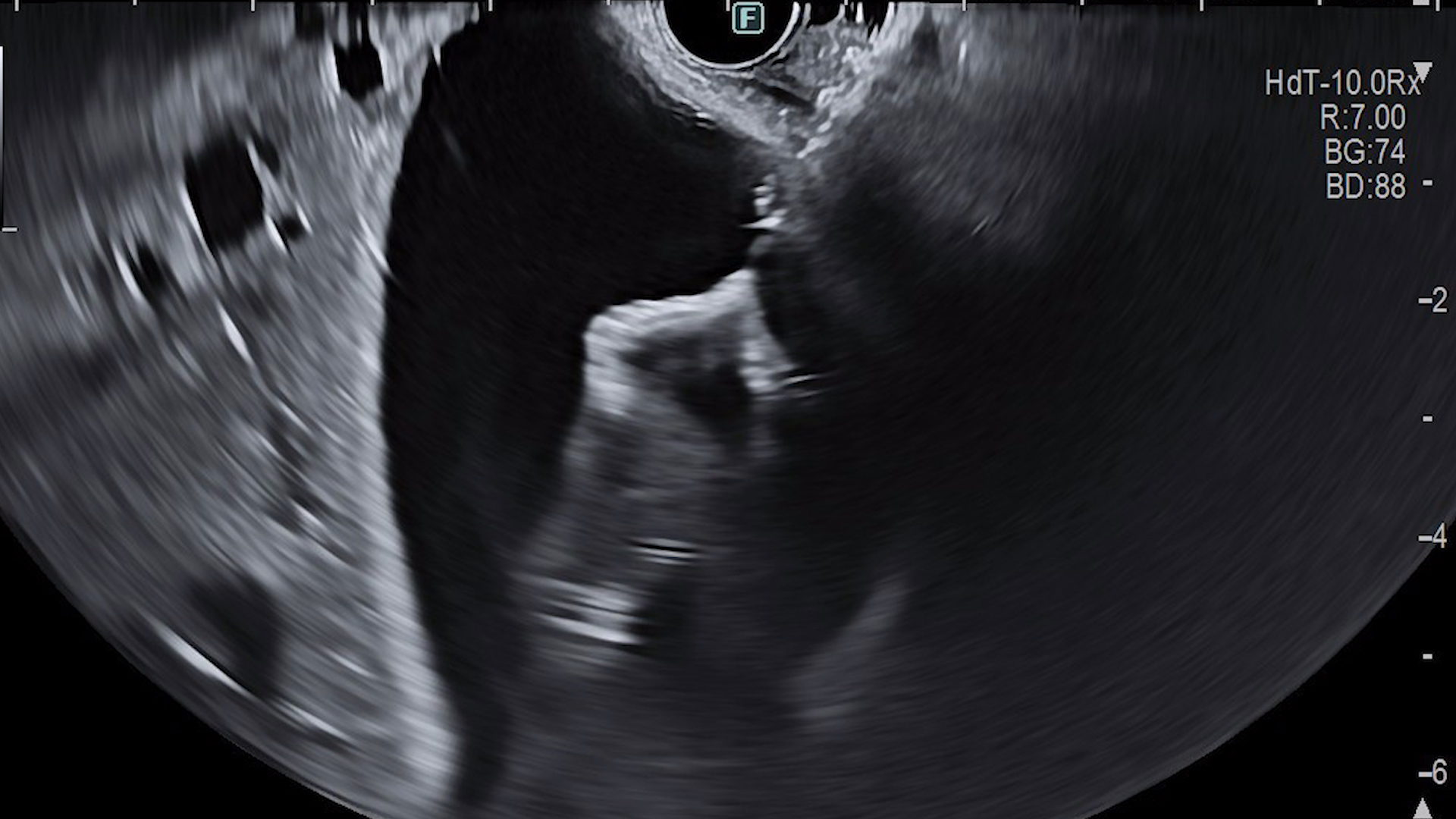
Photo 1 – EUS: large pancreatic collection with heterogeneous content, punctured under EUS guidance.
Photo 2 – Placement of a double-pigtail plastic stent over a guidewire for the initial transmural drainage.
Photo 3 – The insertion catheter crossing the gastric wall at the transmural drainage site.
Photo 4 – EUS: deployment of the lumen-apposing metal stent (Hot AXIOS) into the collection.
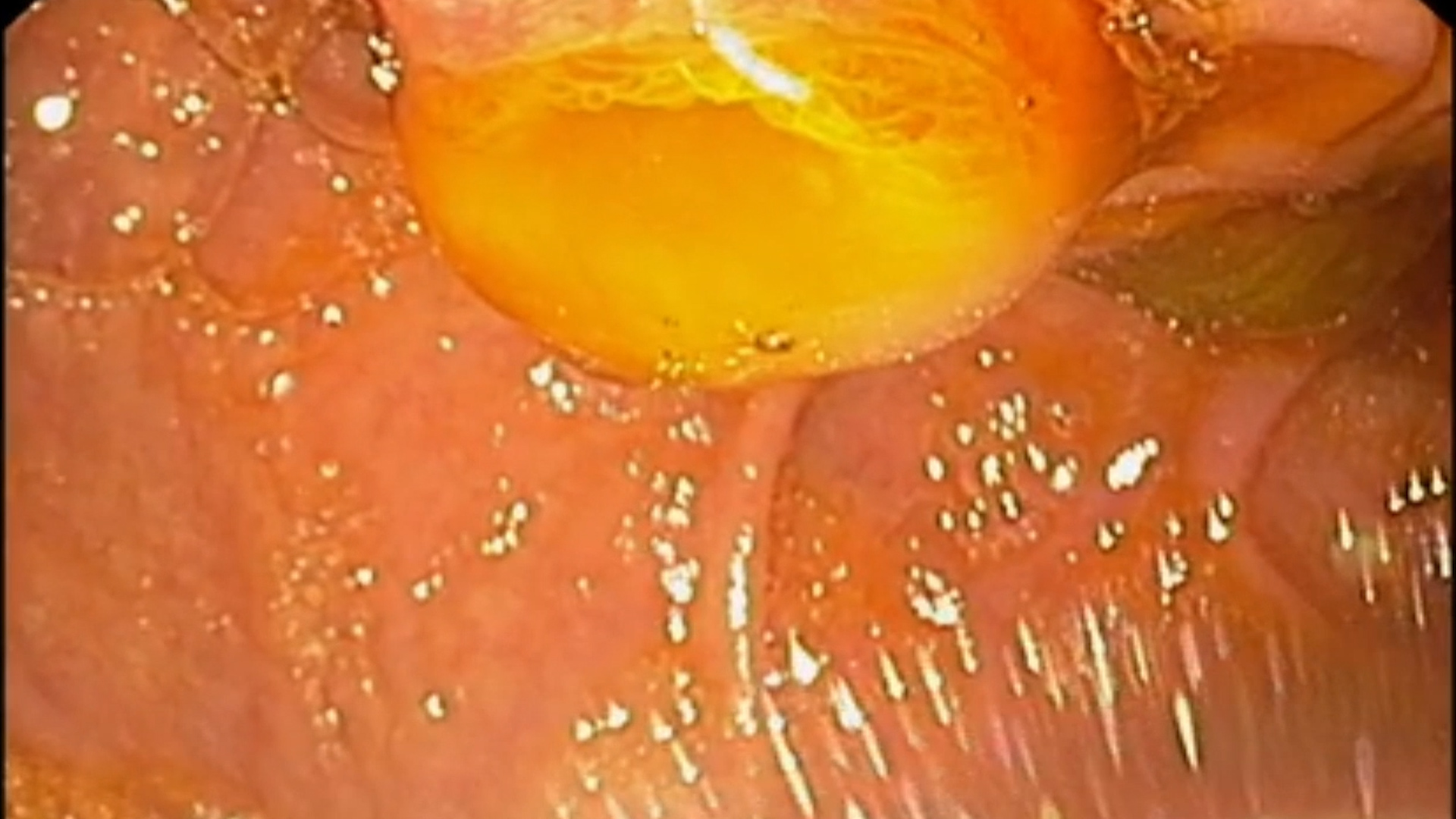
Photo 5 – Gastric flange of the Hot AXIOS stent after deployment.
Photo 6 – View through the stent into the necrotic cavity: necrotic debris and heterogeneous content.
Photo 7 – Endoscopic necrosectomy: necrotic material seen through the stent.
Photo 8 – Cavity with partially evacuated necrotic debris and granulation tissue at a later necrosectomy session.
Photo 9 – Hot AXIOS stent in place, seen from the stomach at endoscopic re-evaluation.
Photo 10 – Clean residual cavity seen through the stent lumen before removal.
Photo 11 – Removal of the Hot AXIOS stent with forceps, four weeks after placement.
Diagnosis
Discussion
The management of pancreatic fluid collections and walled-off necrosis requires an individualized approach based on the clinical presentation, maturity and content of the collection, presence of infection, and response to initial treatment. In this case, the patient initially underwent a minimally invasive step-up approach consisting of EUS-guided transmural drainage with a lumen-apposing metal stent, followed by repeated endoscopic necrosectomy. This resulted in substantial reduction of the collection and initial clinical improvement.
However, shortly after stent removal, the patient developed recurrent fever and abdominal pain, accompanied by a rapid increase in the size of the residual collection from 40 × 53 × 45 mm to 62 × 85 × 58 mm. The heterogeneous content, progressive enlargement and inflammatory changes involving the adjacent gastric wall raised concern for persistent or recurrent infection. Surgical exploration confirmed the presence of a pseudocystic abscess, requiring evacuation and definitive internal drainage.
Endoscopic transmural drainage and endoscopic necrosectomy are currently key components of the minimally invasive management of infected pancreatic necrosis. Nevertheless, failure or recurrence after endoscopic treatment may require escalation to surgical therapy.
In this patient, the laparoscopic/robotic approach allowed evacuation of the infected collection, assessment of the local anatomy and creation of a durable internal drainage pathway through a cystogastric anastomosis. The suspected focal splenic vein thrombosis represented an additional complication requiring careful preoperative assessment. The absence of pseudoaneurysm and the preserved patency of the porto-mesenteric venous system were relevant findings for treatment planning.
This case highlights the importance of close clinical and radiological follow-up after endoscopic drainage and particularly after removal of a lumen-apposing metal stent. Recurrence of symptoms associated with rapid enlargement of a residual collection should prompt early multidisciplinary reassessment.
Conclusions
• Pancreatic collections may recur after apparently successful endoscopic drainage and necrosectomy.
• Recurrent fever, abdominal pain and rapid enlargement of a residual collection after stent removal should raise suspicion of persistent or recurrent infection.
• Endoscopic treatment is an important component of the step-up approach, but surgical intervention remains necessary in selected refractory or complicated cases.
• Laparoscopic and robotic surgery may provide an effective minimally invasive alternative when endoscopic therapy fails to achieve definitive source control.
• Management of complex pancreatic collections should be based on multidisciplinary decision-making.
• Careful imaging follow-up after LAMS removal is essential to identify residual/recurrent collections and vascular complications.
References
- Arvanitakis M, Dumonceau JM, Albert J, et al. Endoscopic management of acute necrotizing pancreatitis: European Society of Gastrointestinal Endoscopy (ESGE) evidence-based multidisciplinary guidelines. Endoscopy. 2018;50:524–546.
- Baron TH, DiMaio CJ, Wang AY, Morgan KA. American Gastroenterological Association Clinical Practice Update on Endoscopic Management of Pancreatic Necrosis. Gastroenterology. 2020;158:67–75.
- van Brunschot S, van Grinsven J, van Santvoort HC, et al. Endoscopic or surgical step-up approach for infected necrotising pancreatitis: a multicentre randomised trial. Lancet. 2018;391:51–58.
- Bang JY, Arnoletti JP, Holt BA, et al. An endoscopic transluminal approach, compared with minimally invasive surgery, reduces complications and costs for patients with necrotizing pancreatitis. Gastroenterology. 2019;156:1027–1040.
- Boxhoorn L, Voermans RP, Bouwense SA, et al. Acute pancreatitis. Lancet. 2020;396:726–734.